

Dr. Giulio A. Santoro, MD, Ph. D, specializes in pelvic floor and colorectal surgery. He is Chief, Tertiary Referral Pelvic Floor and Incontinence Center, Treviso; Director, Italian School of Pelvic Floor Ultrasonography; Associate Professor of Surgery, Italy; and Past-President of the Italian Society of Colorectal Surgery.
Earlier this year Dr. Giulio A. Santoro shared his insights with us about using advanced 3D ultrasound imaging to visualize details of anorectal conditions. One of the main topics in his presentation was anal fistulas. Read on to learn why Dr. Santoro uses 3D ultrasound to prepare for anal fistula surgery.What is an anal fistula?
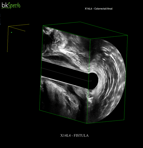
An anal or rectal fistula is a narrow tunnel connection between the rectum/anus and the skin. These are hollow tracks that form inside the body when the anal crypt is obstructed due to bacterial overgrowth which leads to the formation of pus. The fistula serves as a passageway for pus to drain to an external opening. A fistula is caused by an acute inflammation. Mostly, this inflammation process originates from anal glands.
Why use 3D ultrasound imaging to prepare for anal fistula surgery?
High-definition 3D ultrasound imaging helps surgeons visualize the details of conditions like anal fistulas. By using this high-resolution imaging, surgeons can visualize the entire pathway of a fistula. First, a 3D endocavity transducer or 3D anorectal transducer is used to acquire 3D images and datasets using ultrasound. Once the images and datasets are acquired, surgeons can manipulate the datasets, slice through the 3D cube, and angle the different planes to prepare for surgery and make a surgical plan.
Dr. Santoro referenced several clinical studies to support the use of 3D endoanal ultrasound (3D-EAUS) for anal fistula surgery. Overall, the studies noted that 3D-EAUS is a superior technique compared to 2D-EAUS1 and should be routinely used in a clinical setting.2 Using 3D-EAUS may assist surgeons in delineating fistula tract anatomy3 which can result in higher accuracy for determining fistula type and higher accuracy for determining fistula height.4
In summary, medical professionals like Dr. Santoro use 3D-EAUS to prepare for anal fistula surgery because it provides the visual details needed for making surgical plans.
Click here to watch Dr. Santoro’s full webinar about anorectal imaging.
1 Garcés-Albir M, García-Botello SA, Espi A, et al. Three-dimensional endoanal ultrasound for diagnosis of perianal fistulas: Reliable and objective technique. World J Gastrointest Surg. 2016;8(7):513-520. doi:10.4240/wjgs.v8. i7.513
2 Ding JH, Bi LX, Zhao K, et al. Impact of three-dimensional endoanal ultrasound on the outcome of anal fistula surgery: a prospective cohort study. Colorectal Dis. 2015;17(12):1104-1112. doi:10.1111/codi.13108
3 Brillantino, A., Iacobellis, F., Di Sarno, G. et al. Role of tridimensional endoanal ultrasound (3D-EAUS) in the preoperative assessment of perianal sepsis. Int J Colorectal Dis 30, 535–542 (2015). doi:10.1007/s00384-015-2167-0
4 Kołodziejczak, M., Santoro, G.A., Obcowska, A., Lorenc, Z., Mańczak, M. and Sudoł-Szopińska, I. (2017), Three-dimensional endoanal ultrasound is accurate and reproducible in determining type and height of anal fistulas. Colorectal Dis, 19: 378-384. doi:10.1111/codi.13580