
Recently, we spoke with Dr. Jos J. Immerzeel, the radiation oncologist at Andros Mannenkliniek (The Andros Men’s Health Institutes) in Amsterdam. Dr. Immerzeel uses bkFusion to plan and guide transperineal prostate biopsies using local anesthesia, which you can read about here.
For Dr. Immerzeel, an MR-ultrasound fusion biopsy using bkFusion takes approximately 20 minutes, and patients usually spend less than an hour at the clinic. One reason for the short procedure time is that the MRI contours have been done beforehand, so the patient spends very little time on the table.
Dr. Immerzeel talked us through two cases where the ultrasound images on the bk3000 showed important new information that was not visible on the MR images.Patient One – Expanding the Contour
This patient was 60 years old and weighed 74 kg (163 lbs). The patient’s MRI data showed a small lesion of 8 mm on the border of the anterior zone and the transition zone, and tests indicated PIRADS 3 and a PSA level of 7. Two years earlier, the same patient was found to have only PIRADS 2. The lesion was not large, and Dr. Immerzeel planned to take systematic biopsies as well as targeted biopsies from the lesion area.
He began the procedure by administering local anesthetic. Dr. Immerzeel injected the anesthesia into the prostate capsule under ultrasound guidance using the grid and biplane transducer (see Fig. 1). He continued until the patient could no longer feel the injections.
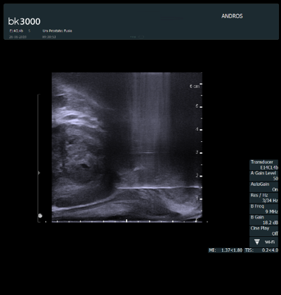 Fig 1. The needle right at the capsule of the prostate ready to insert the anesthesia. Sagittal view.
As the biplane transducer was already inserted and the grid in place, Dr. Immerzeel could immediately begin fusing the ultrasound image with the MR images.
bk3000 Shows More
The real-time ultrasound images on the bk3000 provided new information during the procedure. The ultrasound images showed a more hypo-dense area, which had not been as clear on the MRI images (See Fig. 2). Dr. Immerzeel used this new information to update his biopsy plan.
Based on the real-time ultrasound images, Dr. Immerzeel widened the lesion contour. He then proceeded to do the targeted biopsies within this newly expanded contour, all without any disruption to his familiar workflow. Without high-resolution ultrasound guidance, it might have been more difficult to see the true size of the lesion (2 of the 3 biopsies were GS3+3).
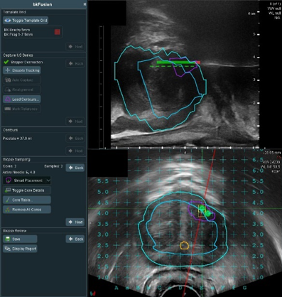 Fig 2. Hypo-density lesion with a registered core. Sagittal and transverse view.
Patient Two – Another Lesion
The second patient was a 64-year-old man, weighing 89.9 kg (198 lbs). The patient’s tests had shown PIRADS 4 with a lesion in the lateral posterior left section of the prostate.
While scanning and fusing the MR and ultrasound images, Dr. Immerzeel discovered what seemed to be a second lesion in a different part of the prostate, more anterior.
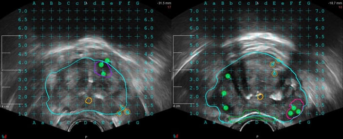 Fig 3. Low echogenic anterior lesion with three registered cores and a lesion in the peripheral zone with three registered cores. Transverse view.
Reviewing the real-time ultrasound images, they showed clearly that there was indeed another lesion (See Fig. 3). He then made new contours on the fused image. With the updated contours, Dr. Immerzeel was able to take targeted biopsies from both lesions and both were GS3+4.
The Importance of Ultrasound in Prostate Biopsy Procedures
These two patient stories show how high-definition ultrasound images on the bk3000 contribute important information to biopsy procedures. By adding this new information to the MRI images, Dr. Immerzeel can make sure that he takes biopsy samples from each region of interest.
The Predictive Fusion® technology adjusts the MR images in preparation for the real-time ultrasound fusion before the patient is even on the table. The high-quality ultrasound images on the bk3000 are then seamlessly overlaid onto the MRI data, helping urologists perform prostate biopsies with ease, whether they do TR or TP. The BK system makes it simple to update prostate biopsy plans – all within the familiar biopsy workflow.