
Pelvic floor disorders lead to hundreds of thousands of surgical interventions on female patients in the USA each year. An estimated 30% of these cases require re-operations.
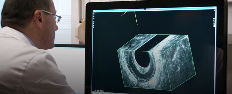
Next generation 3D ultrasound imaging is helping physicians be more effective in treating these disorders. It enables practitioners, in real time, to see better and to better determine the right treatment or surgical intervention.
As Dr. Santoro from the Department of Surgery at the Regional Hospital in Treviso, Italy, and an expert in pelvic floor disorders says, “3D ultrasonography provides better insight into the causal mechanisms underlying clinical presentation and improves surgical technique.”
The Advantages are Easy to See
During our discussions with Dr. Santoro, he noted that the pelvic floor is very complex. “It comprises different compartments, muscles, ligaments and nerves. It is not only anatomy. It is also functional. 3D diagnostic ultrasound provides detailed, dynamic images of the entire pelvic floor.”
In many ways, Santoro’s staff mirrors the complexity of the pelvic floor. “We have gastroenterologists, urologists, radiologists and physical therapists in our unit because we think that the approach to these patients should be multi-disciplinary,” he says.
Three-dimensional ultrasonography allows for multiplanar imaging of both rectal and anal tumors that would normally require the use of expensive MRI systems. “Of course, MRI is very sophisticated,” Dr. Santoro says, “but 3D ultrasonograhy is easier and more comfortable for the patient and we can easily use it in our offices.”
The latest generation of 3D imaging greatly improves visualization of surrounding structures and organs, giving clinicians a more complete and integrated picture of the problem. It also enables active patient participation during the assessment. Practitioners can actually see what happens when the patient is asked, for example, to squeeze.
3D ultrasound technologies can provide information that can help doctors make an earlier diagnosis and decide on treatment of pelvic floor disorders, including rectal and anal tumors, which require early intervention for increased rates of success.
For clinics and hospitals, 3D ultrasound offers major workflow time-savings. Physicians get results almost immediately. “We can reproduce results with 3D ultrasound”, Dr. Santoro emphasized, “This is important. We can do this procedure in the operating room pre-operatively. It helps guide the physician in choosing the best approach.”
As a bonus, 3D ultrasound’s relative price point makes seeing into the body and determining the appropriateness of various surgical alternatives an affordable imaging tool.