
When performing a laparoscopic cholecystectomy, intraoperative ultrasound (iUS) is a good alternative to intraoperative cholangiography because it provides a fast, safe, and repeatable means of visualizing the procedure, with clear details and soft tissue delineation. Read on to learn more about the uses and benefits of laparoscopic intraoperative ultrasound when compared with other methods.
Unlike intraoperative cholangiography, iUS is non-radiating, which helps reduce radiation hazards to patients and staff. iUS has also been shown to be completed more rapidly than IOC, with one study reporting a laparoscopic ultrasound duration of 9.8 minutes versus 17.6 minutes for IOC.1
Why Is iUS Valuable?
The list below explains some of the fundamental reasons why iUS is valuable in cholecystectomy procedures.
- Get real-time images as often as necessary throughout the procedure. Imaging with intraoperative ultrasound can be repeated as many times as desired during cholecystectomies.
- Use existing ports. Another value to using iUS in cholecystectomies is that iUS can be performed using the existing ports in a standard 4-port cholecystectomy. Laparoscopic ultrasound transducers such as the 4-way flexible Advanced Laparoscopic Transducer from BK Medical can be placed through the epigastric or umbilical port.2, 3
- Help visualize the anatomy during the procedure. iUS helps visualize the variations, vascularity, and the anatomical structure beyond the visible surface. By helping to visualize anatomy during the procedure, iUS helps in avoiding misidentification.
- Analyze common bile duct. iUS can be used to assess and confirm the integrity of the common bile duct (CBD).
Steps Where Ultrasound Is Used in the Procedure
iUS can be used in every step of the cholecystectomy procedure to help support surgical decision-making.
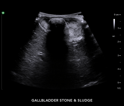
- Staging: Screen and confirm for the presence of common bile duct stones at the beginning of the procedure. Real-time intraoperative ultrasound supports a thorough inspection of the gallbladder that also helps surgeons understand the presence of gallbladder polyps (if any) and differentiates between the thickened walls and sludge.4
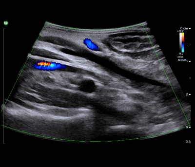
- Planning and Intervention: Leverage real-time information for planning and guiding the procedure. Laparoscopic ultrasound allows visualization of anatomical variations of the biliary tree beyond the surface and helps delineate the anatomical and vascular relationships. Visualizing and understanding these variations and relationships is critical to safe dissection. Surgeons can understand vascular relationships by outlining the portal vein, CBD, and hepatic artery, as well as visualizing the intrapancreatic portion of the CBD.
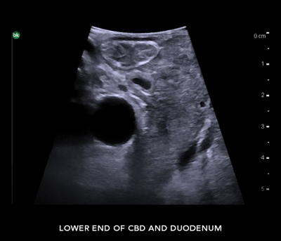
- Validation: Post-dissection, utilize intraoperative imaging to assess for completeness of the resection and trace the CBD to its exit point into the second part of the duodenum.
Overall, using intraoperative ultrasound for cholecystectomy procedures gives you the ability to visualize and evaluate biliary and vascular anatomy.
Learn more about bkActiv and active imaging with intraoperative ultrasound.
1 Dili A, Bertrand C. Laparoscopic ultrasonography as an alternative to intraoperative cholangiography during laparoscopic cholecystectomy. World journal of gastroenterology. https://pubmed.ncbi.nlm.nih.gov/28839445/. Published August 7, 2017.
3 I13C3f has not been licensed by Health Canada