Dr. Robert Martin is a professor of surgery at the University of Louisville School of Medicine in Louisville, Kentucky, and the division director of surgical oncology. His background is in surgical oncology, with additional training in hepatopancreatobiliary surgery. Most of his practice today is spent treating patients with upper gastrointestinal cancer. His management of this disease always includes the use of ultrasound.
The first area where Dr. Martin finds ultrasound essential to managing patients with upper gastrointestinal cancers is in staging. It helps him determine if a patient’s disease is localized or metastatic. The second situation is in two areas where cross-sectional imaging has its underlying limitations: in a hilar or perihilar cholangiocarcinoma and in a pancreatic head tumor. In both of these areas, a lot of volume averaging occurs with a CT scan or MRI because of multiple vital structures that are in proximity. In CT or MRI, volume averaging is the effect of expressing the average density of a voxel as a pixel in the image; the greater the slice thickness, the more averaging is necessary, with loss in density resolution. Based on only a CT or MRI scan, the surgeon may think that a tumor is resectable or marginally resectable. Intraoperative ultrasound can then be used as an aid to either confirm that the disease is not too extensively advanced and that the patient can undergo surgical resection; or that the disease is more advanced or more extensive locally than was previously seen, and the surgeon should not try to perform an extensive dissection.
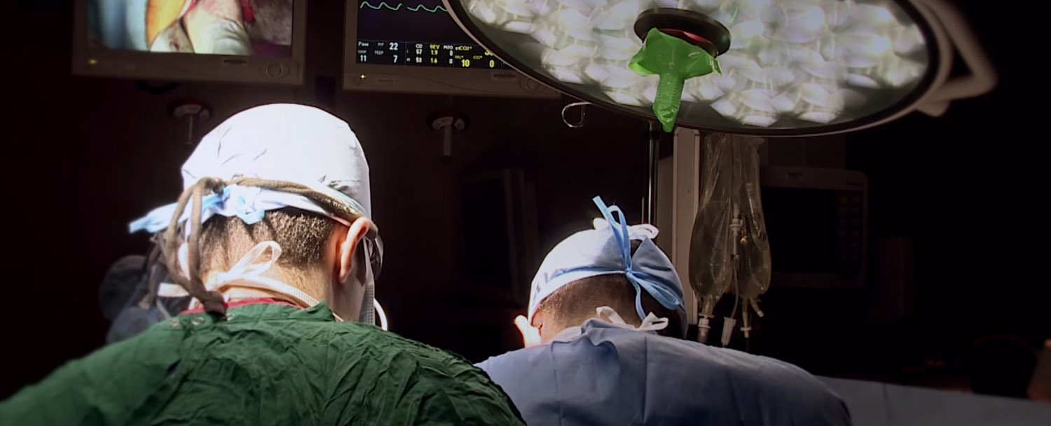
"Using intraoperative ultrasound for any of my cases is now just as important as suture or any kind of dissecting device,” Dr. Martin explained. “It’s an additional surgical instrument that I think is vitally important. It’s quick, inexpensive, and not harmful to the patient.” Ultrasound can be used to help with partial gastrectomy for a GIST, evaluate an enucleation for a liver cyst, or help determine whether or not to resect a hemangioma. “I can give you multiple reasons why ultrasound is going to help you in your diagnostic accuracy and your surgical quality,” he continued. “If you’re doing anything in the upper gastrointestinal area, whether it’s benign or malignant, I can guarantee there’s a clear reason to use ultrasound in some way.”
Dr. Martin has been mainly using BK Ultrasound systems for almost nine years because they address the needs of the surgeon. He has found the BK 8666-RF laparoscopic transducer particularly effective when working with obese patients. The length of the transducer allows him to reach across the patient, and the 4-way manipulation gives him the flexibility to reach difficult-to-access areas, reducing frustration in the operating room. For the majority of his pancreatic and liver ablations, Dr. Martin uses the BK biplane probe. Being able to image in two dimensions in real time is important for accuracy and needle placement, and helps with patient safety and efficacy when using any type of ablative therapy.
In the management of upper gastrointestinal diseases, great advances have been made in the collaborative multidisciplinary approach. Dr. Martin and his team have been focused on innovative ablative devices, either thermal-based ablative devices — predominantly microwave ablation — or non-thermal ablative devices, that being irreversible electroporation. Another area of great interest to Dr. Martin is regional therapy, which is the delivery of high-dose radiation or chemotherapy directly to the target organ. In this way, the patient avoids the side effects from systemic chemotherapy, which can adversely affect the whole body. “Regional delivered therapies can potentially be adjunctive to systemic chemotherapy,” Dr. Martin said. “Not as a replacement, but to help enhance the response rate. A lot of my focus has been on optimizing that type of regional delivery therapy, especially in the liver.”1
Dr. Martin is also focused on optimizing endoscopic management of disease, especially with regard to patients who have premalignant lesions. When there is a small premalignant lesion, he uses focal endoscopic or ablative therapies to eradicate the lesion rather than removing the entire organ. In this way, he can help reduce the overall surgical morbidity, potentially spare the patient from developing a more invasive cancer, or subject the patient to more intensive types of therapy.
Given the growing number of treatment options available today, Dr. Martin advises patients who are diagnosed with an upper gastrointestinal cancer to become empowered by learning as much as they can right from the start so they understand all of their options.
Visit our surgery page to learn more about how our ultrasound systems can be used for intraopertive procedures. Click here to learn more about our new bk5000 surgical ultrasound system.
1 Farlex Partner Medical Dictionary. S.v. “volume averaging.” Retrieved August 7 2015
from http://medical-dictionary.thefreedictionary.com/volume+averaging

